Easy Drawing of a Cell Membrane

Cell membrane in a cell easy drawing – Let’s embark on a journey into the microscopic world, where we’ll learn to depict the cell membrane—that remarkable boundary that keeps a cell’s inner world separate and safe. Drawing the cell membrane can seem daunting, but with a few simple steps, you can create a visually appealing and informative diagram that captures its essence. This isn’t just about lines and shapes; it’s about understanding the fundamental structure and function of this crucial cellular component.
Understanding the cell membrane’s structure is key to drawing it accurately. Think of it as a dynamic, bustling city, with various components moving and interacting. Our drawing will simplify this complexity, focusing on the essential features.
Simplified Cell Membrane Drawing Steps
The following steps will guide you through creating a simplified, yet informative, drawing of a cell membrane. Remember, the goal is to illustrate the key components and their arrangement, not to create a photorealistic image.
The fragile cell membrane, a simple sketch, a whispered secret of life’s delicate dance. Its permeable boundary, a shield against the vast unknown, reminds me of the stark lines in a drawing of soldiers, a similar simplicity in representation; find inspiration for those lines at army men drawing easy , then return to the quiet beauty of the cell membrane, a microcosm of resilience, drawn with a gentle hand.
- Draw a rectangle: Begin by drawing a long, thin rectangle to represent a small section of the cell membrane. The proportions aren’t critical; focus on making it easily visible.
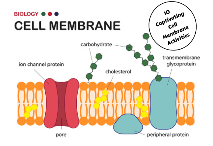
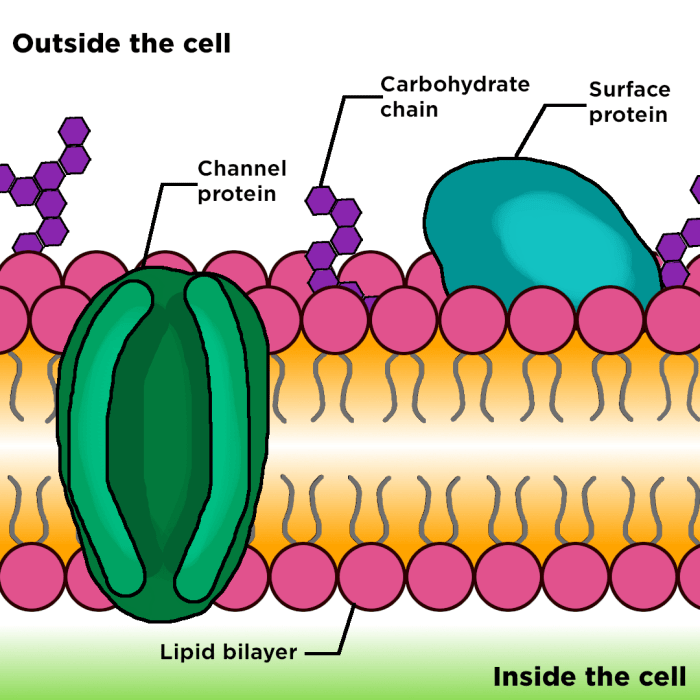
- Sketch the phospholipid bilayer: Within the rectangle, draw two parallel lines, slightly wavy, to represent the hydrophilic heads of the phospholipid molecules. These heads are attracted to water, so they face outward, toward the cell’s interior and exterior. Between these lines, draw a slightly thicker, less defined space representing the hydrophobic tails of the phospholipids. These tails repel water and hide inside the membrane.
- Add proteins: Scatter a few different shapes throughout the phospholipid bilayer. Some proteins should span the entire membrane (transmembrane proteins), while others should be embedded within only one layer. These shapes represent the diverse proteins within the membrane, each with a unique function, such as transport or signaling.
- Include carbohydrates: Add small, branched shapes attached to the outer surface of the phospholipid heads. These represent carbohydrate chains, often attached to proteins (glycoproteins) or lipids (glycolipids). These play a crucial role in cell recognition and communication.
- Label the components: Clearly label each component: phospholipid bilayer (heads and tails), transmembrane protein, embedded protein, carbohydrate. Use concise labels and a clear font.
Tips for a Visually Appealing Diagram
Creating a visually appealing diagram enhances understanding. A well-executed drawing makes the complex structure of the cell membrane more accessible and memorable.
- Use contrasting colors to differentiate the phospholipid bilayer components (heads and tails), proteins, and carbohydrates. For instance, use a darker shade for the hydrophobic tails and a lighter shade for the hydrophilic heads.
- Maintain a consistent scale. While not perfectly to scale, keep the relative sizes of the components reasonably proportional.
- Use clear, concise labels. Avoid overcrowding the diagram with excessive text.
- Keep the lines neat and clean. A well-drawn diagram is more aesthetically pleasing and easier to understand.
Microscopic Appearance of the Cell Membrane
Under a powerful microscope, the cell membrane appears as a thin, dark line separating the cell’s interior from its surroundings. The individual components—phospholipids, proteins, and carbohydrates—are too small to be resolved with standard light microscopy. However, the membrane’s overall structure and its interactions with other cellular components can be inferred from microscopic images, often using techniques like electron microscopy, which provide much higher resolution.
For example, techniques like freeze-fracture electron microscopy reveal the presence of embedded proteins within the membrane as bumps and pits on the fractured surfaces. These observations support the fluid mosaic model, which describes the cell membrane as a dynamic, fluid structure with various components moving within it.
Cell Membrane Interactions: Cell Membrane In A Cell Easy Drawing

The cell membrane, that breathtakingly delicate yet incredibly robust barrier, isn’t just a passive wall separating the inside of a cell from the outside world. It’s a dynamic interface, a bustling crossroads where life’s essential transactions occur. It’s a place of constant interaction, a silent symphony of molecular exchanges that orchestrate the cell’s very existence. Understanding these interactions is key to understanding life itself.The cell membrane interacts with its environment in a myriad of ways, constantly adapting and responding to external stimuli.
It’s a selectively permeable barrier, carefully choosing what enters and exits the cell, maintaining the delicate internal balance crucial for survival. This selective permeability is achieved through a complex interplay of lipids, proteins, and carbohydrates embedded within the membrane’s fluid mosaic structure. Think of it as a sophisticated gatekeeper, allowing nutrients in and waste products out, all while protecting the cell’s precious cargo.
Receptor Proteins in Cell Signaling
Receptor proteins, embedded within the cell membrane, act as the cell’s sensory organs. These specialized proteins bind to specific signaling molecules, such as hormones or neurotransmitters, triggering a cascade of events within the cell. Imagine these receptors as tiny locks, each with its own unique key. When the right key (signaling molecule) arrives, the lock opens, initiating a chain reaction that alters the cell’s behavior – perhaps triggering growth, initiating a metabolic process, or even triggering cell death.
This intricate communication system allows cells to coordinate their activities and respond to changes in their environment, ensuring the organism’s overall well-being. For instance, insulin binding to its receptor on muscle cells triggers glucose uptake, regulating blood sugar levels.
Cell Junctions and Their Functions
Cells don’t exist in isolation; they’re part of a larger community, often interacting closely with their neighbors. Cell junctions are specialized structures that connect adjacent cells, providing structural support and facilitating communication. Tight junctions, for example, create a watertight seal between cells, preventing leakage between them. This is crucial in tissues like the lining of the intestines, where it prevents harmful substances from entering the bloodstream.
Gap junctions, on the other hand, create channels that directly connect the cytoplasm of adjacent cells, allowing for rapid communication and the exchange of small molecules. This is essential in tissues like heart muscle, where coordinated contractions are vital for efficient pumping. Finally, adherens junctions and desmosomes provide strong mechanical attachments between cells, contributing to the structural integrity of tissues.
Endocytosis and Exocytosis, Cell membrane in a cell easy drawing
The cell membrane isn’t just a static barrier; it’s also incredibly dynamic, constantly reshaping itself to accommodate the import and export of materials. Endocytosis is the process by which the cell engulfs extracellular material, forming vesicles that bring the material into the cell. This can be through phagocytosis (cell eating), pinocytosis (cell drinking), or receptor-mediated endocytosis (a more targeted form of uptake).
Imagine the cell membrane extending outwards, wrapping around a particle like a hungry amoeba, and then pinching off to form a vesicle carrying its prize inside. Exocytosis, conversely, is the process by which the cell expels materials from its interior. Vesicles containing these materials fuse with the cell membrane, releasing their contents into the extracellular space. This is how cells secrete hormones, neurotransmitters, and other essential substances.
Think of it as the reverse of endocytosis, a vesicle merging with the membrane and releasing its cargo into the environment. These two processes are essential for cell communication, nutrient uptake, and waste removal.
Passive Transport Mechanisms
Passive transport mechanisms move substances across the cell membrane without requiring energy input from the cell. These processes rely on the inherent properties of the membrane and the concentration gradients of the substances being transported.
- Simple Diffusion: Movement of a substance from an area of high concentration to an area of low concentration, directly across the lipid bilayer. Think of a drop of dye spreading out in a glass of water.
- Facilitated Diffusion: Movement of a substance across the membrane with the assistance of membrane proteins, still following a concentration gradient. This is like using a doorway to cross a wall instead of climbing over it.
- Osmosis: Movement of water across a selectively permeable membrane from an area of high water concentration to an area of low water concentration. This is crucial for maintaining cell volume and turgor pressure.
Questions and Answers
What are some common diseases related to cell membrane dysfunction?
Defects in cell membrane structure or function can lead to various diseases, including cystic fibrosis (due to faulty chloride channels), muscular dystrophy (affecting membrane proteins in muscle cells), and certain types of cancer (due to altered membrane receptors).
How does the cell membrane contribute to cell signaling?
Receptor proteins embedded in the cell membrane bind to specific signaling molecules (ligands), triggering intracellular signaling cascades that influence cell behavior and function.
Can you explain the difference between integral and peripheral membrane proteins?
Integral proteins are embedded within the lipid bilayer, while peripheral proteins are loosely associated with the membrane surface, often interacting with integral proteins.
Why is cholesterol important in the cell membrane?
Cholesterol helps maintain membrane fluidity by preventing the phospholipids from packing too tightly together at lower temperatures and preventing them from becoming too fluid at higher temperatures.
