Image Description and Analysis: Animal Cell Coloring Page.jpg

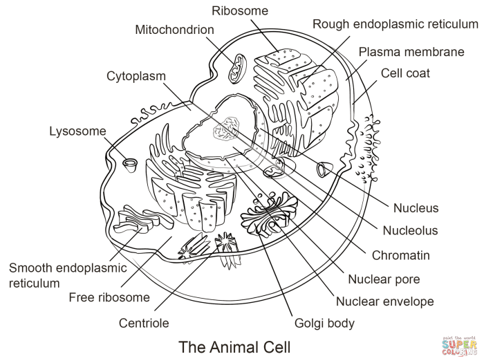
Animal cell coloring page.jpg – The animal cell coloring page presents a simplified, cartoonish representation of an animal cell, likely intended for educational purposes, particularly for younger learners. The overall style is bright and playful, employing bold Artikels and large, easily identifiable organelles. The color palette is vibrant, using primary colors and some secondary shades, making it visually appealing and engaging for children.The image depicts several key organelles, although not all are labeled.
The nucleus is centrally located and significantly larger than other organelles, depicted as a large, round shape, likely colored in a light shade such as pale yellow or light pink. Several smaller, oval-shaped mitochondria are scattered throughout the cytoplasm, potentially in a reddish-brown or dark purple hue. Ribosomes are suggested, possibly represented as small dots, although their individual identification is difficult without labeling or coloring.
The cell membrane is a simple Artikel, surrounding all other components. The cytoplasm itself may be a light blue or pale green. There is no depiction of more complex organelles like the Golgi apparatus, endoplasmic reticulum, or lysosomes.
Organelle Representation and Spatial Arrangement, Animal cell coloring page.jpg
The relative sizes and positions of the organelles are simplified for clarity. The nucleus is disproportionately large compared to its actual size relative to the rest of the cell. The mitochondria are numerous but their arrangement doesn’t reflect their dynamic nature within a real cell. The absence of many other important organelles, such as the endoplasmic reticulum and Golgi apparatus, significantly simplifies the overall representation.
The spatial distribution of the organelles, while visually pleasing, lacks the detailed complexity found in a scientifically accurate diagram.
Comparison to Scientific Accuracy
This coloring page offers a vastly simplified representation compared to a scientifically accurate diagram of an animal cell. A scientifically accurate diagram would show the intricate internal structure, including the endoplasmic reticulum (a network of membranes), the Golgi apparatus (a stack of flattened sacs), and lysosomes (small, membrane-bound organelles). The relative sizes and spatial arrangements of the organelles would be more precise.
For instance, the mitochondria would be smaller and more numerous, and the nucleus would occupy a proportionally smaller space. Furthermore, a scientifically accurate diagram might include details of the cytoskeleton, which provides structural support to the cell, and is absent in this simplified version.
Caption Suggestion
“Explore the amazing world inside an animal cell! Color this page to learn about the different parts and their functions.”
Right, so you’re into that animal cell coloring page.jpg, proper sciencey vibe, innit? But if you fancy something a bit cuter, check out these baby animal coloring pages free printable – they’re dead cute! Then, after you’ve had a chill sesh with the fluffy critters, you can get back to your wicked animal cell coloring page.jpg and show off your artistic skills, yeah?
